The most basic breakdown of the ear is into 4 parts: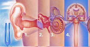
- The OUTER EAR
- The MIDDLE EAR
- The INNER EAR
- The AUDITORY NERVOUS SYSTEM (ANS)
Outer Ear
The outer ear consists of two parts—the pinna and the external auditory canal (ear canal). The most visible and easily recognized (as well a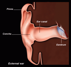 s adorned by jewelry) part of the ear is the pinna (also called the auricle) which is flap of cartilage covered by skin. The pinna is uniquely shaped with many superficial bumps and grooves to help funnel sound to the ear canal while providing subtle “filtering” clues which help us know the direction a sound is coming from (localization).
The external auditory canal (ear canal) continues to funnel sound deeper to the tympanic membrane (ear drum) over a distance of a little over one inch (25-28 mm). The underlying structure of the canal is approximately 1/3 (outer portion) cartilage and 2/3 (deeper portion) bone which is covered in thin tissue/skin. Also, primarily in the outer third of the canal, we have cerumen (ear wax) producing glands and many tiny hair follicles which serve as protection for the deeper, more delicate structures of the middle and inner ear.
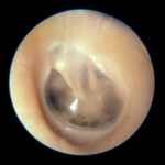
This protective system traps dust/debris, waterproofs the ear, and helps prevent harmful bacteria and fungi from “setting up shop” and causing external ear infections (e.g., “swimmer’s ear”). Most people do not realize that this system is also relatively self-cleaning as new skin growth pushes old tissue (along with old wax/debris) outward to the opening of the canal at the pinna at approximately 0.1 mm per day (about the same speed as fingernail growth). The debris then dries and washes/flakes away during normal bathing/activities.
Finally, the physical dimensions of the outer ear allow for some natural amplification. The funneling and resonating effects of the pinna and external canal adds approximately 10-15 decibels in the 1.5 to 7 kHz frequency range (mid to high pitches), which happen to be in the range of much important human speech information.
Middle Ear
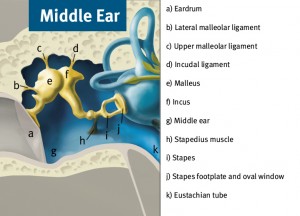 The space between tympanic membrane (eardrum) and the outer boundary of the inner ear (called the promontory) is referred to as the middle ear. The middle ear space is approximately 2 cubic centimeters in volume and is connected to the nasopharynx (nose/throat cavity) by a long tube called the eustachian tube. This tube allows for equalization of air pressure differences across the tympanic membrane (match air pressure of the middle ear space to that of outside air) by opening for a short time while yawning or swallowing. Many people hear this opening of the eustachian tube and air pressure equalization as a “snap”,”crackle”, or “pop” (reminds me of a popular breakfast cereal). Loss of proper eustachian tube function can lead to a muffled sense of hearing, ear pain, and/or fluid build up behind the eardrum (may or may not be infected). See the section on conductive hearing loss for more on this.
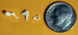 The primary anatomy of the middle ear consists of the tympanic membrane, the middle ear ossicles (bones), and the eustachian tube. The ossiclular chain is comprised of three bones which connected in order from the eardrum to the inner ear starting with the malleus (“hammer”), followed by the incus (“anvil”), and finally the stapes (“stirrup”). The stapes is the smallest bone in the human body and attaches to a very small membrane opening to the inner ear, called the oval window.
Our middle ear systems are designed to convert (or “transduce”) sound vibrations in the air into mechanical energy which is then passed on to the inner ear fluid. The eardrum is approximately 17 times larger than the oval window which results in an increase in pressure as the mechanical energy is passed on to the inner ear. Along with other mechanical advantages (think of a lever) of the ossicular chain, this system results in a maximum total pressure increase of 44 times of the original sound pressure captured at the eardrum to the stapes footplate at the oval window (corresponds to 33 decibels of hearing).
Trivia: the smallest muscle in our body is also located in the middle ear, which is called the stapedial muscle. This muscle is one of the several ligaments and muscles that suspend the ossicular chain within the middle ear space. The stapedius along with the tensor tympani muscle not only help hold the ossicles in place, but contribute to an acoustic reflex. The reflex triggers a stronger contraction of the muscles in response to loud sounds (over 80 decibles) and dampens ossicular vibration, therefore reducing sound transmission by as much as 10 to 30 decibels. This is believed to be either protective in nature for sudden and brief loud sounds and/or to help reduce sound distortion as a result of excess vibration. The acoustic reflex can be measured by audiologists as part of diagnostic testing since the reflex relies on proper function of the ear, hearing nerve, brainstem, and facial nerve (nerve to the muscle) which helps to determine the location/site of a problem. In fact, muscle contraction can be measured on the ear opposite to the ear in which the headphone delivered the loud tone. Got all that? Now go wow your friends!
Inner Ear
The inner ear is where all the “magic” happens—or in other words, where vibration is sorted and turned into signals that our brain can understand. Throughout this section, as I explain how this amazing process unfolds, please remember that I am still telling a very simplified story and the true complexities of the inner ear (both known and yet unknown) could fill quite a large volume of pages.
The inner ear is housed in the temporal bone of the skull and is really comprised of several canals or tunnels through the bone—in fact, this is often referred to as the bony labyrinth (labyrinth meaning “maze”). Inside of the bony labyrinth is soft structure which is referred to as the membranous labyrinth, on which the very important cellular and sensory structures are located. All other space is filled with fluid (remember that I mentioned “hydraulic energy” in the previous section).
The inner ear is typically discussed as having three main parts—the cochlea, vestibule, and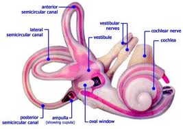 semicircular canals. While these parts are separated by function, they are actually all interconnected with each other. Under normal circumstances, the vestibule and semicircular canals are involved in our sense of balance (rather than hearing) and will not be discussed in this section. The hearing part of our inner ear is the cochlea, which is coiled in such a way that it looks very similar to a snail’s shell and is about the size of a pea. This coiled tube is divided (length-wise) by 2 membranes into 3 compartments. The middle compartment is called the scala media and contains the most important structures for detecting and organizing sound vibrations which have passed from the middle ear into the fluid of the inner ear (from the stapes through a membrane called the oval window).
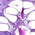
The best way to conceptualize the workings of the cochlea is to think of it uncoiled (straightened out). By nature and arrangement of cells and the properties of the basilar membrane that they are built upon, this uncoiled cochlea functions much like a xylophone. By this, I mean that at one end the basilar membrane is narrow and stiff and the other end is wide and more flaccid (loose). As sounds enter the fluid of the inner ear, they naturally “find” the place along this structure that matches in “tune” (resonates). The structures and haircells upon the membrane at those particular locations are then stimulated—this one major way complex sounds are organized for our nerve and brain (the other has to do with the timing of nerve pulses that follows the vibration pattern).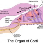
Haircells are contained within a specialized mechanical structure called the organ of corti. Haircells are THE sensory cells of the inner ear and are named so by their appearance—they are cylinder-shaped cells with “hairs” standing on top, called cilia or stereocilia. When stereocilia are “bent” back and forth from a sound vibration, the cell is activated. We have around 40 to 150 stereocilia on each haircell and approximately 30,000 haircells between our two ears. Only about one quarter to one third of these haircells actually send sound information to our nerve and brain when activated—these are called inner haircells. All of our other haircells in the cochlea are called outer haircells and are responsible for increasing tuning and sensitivity (they are often referred to as “cochlear amplifiers”). Outer haircells are special in that exhibit motility when activated. In other words, they MOVE!
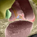
More precisely, they get longer and shorter in synchrony with the incoming vibration to help enhance stimulation of the inner haircells. I like to picture the inner haircells as your younger brother swinging (vibratory movement) on a swing-set. You are the older sibling (outer haircells) giving an “extra push” from behind each time he comes back to you. In doing so, energy is added to the system and your brother swings higher! Without outer haircells, we would lose approximately 50 decibels of hearing sensitivity!
Haircells are in direct communication (using tiny molecules called neurotransmitters) with tiny individual fibers of the auditory nerve (Cranial Nerve VIII), which convert the chemical message into neural impulses and forward the information to (and from) our brains. The high to low frequency (pitch) information that was sorted by the basilar membrane and haircells is preserved by the organization of nerve fibers and centers of the brainstem and brain. More on this in the next section Auditory Nervous System.
Trivia: The cochlea is fully formed and functional by 30 weeks of gestation (near the beginning of the 3rd trimester of pregnancy) and remains the same size for one’s entire life.
Auditory Nervous System
Coming soon! (this section has nothing to do with your ear’s lack of confidence!)
Connect